vendredi 18 juillet 2014

Let's talk about inhibition !!!


Topic pretty complex that will be treated in several posts.


Inhibition was already discussed previously in « world of fluorophores part 2 » post about most used molecules and the way to give a sample result considering inhibition control results.


Nevertheless, here I will talk about how to characterize inhibition from PCR results but also to get rid of inhibition more or less and how to give a result considering inhibition results.


Before any deeper analysis of this topic, a good thing is to know whether your PCR runs have been even slightly subject to inhibition. Few hints can help you : 


-         -  Imperfect sigmoid amplification signal with :

o   Curve flattening

o   Low raw flurorescence level

o   Abnormal shape of amplification curve

o   Exponential phase of the curve widened or flattened

- 
       -   Dilution non recovered by PCR results (eg. If you have less than 3.32 Ct between pure and 1/10 diluted sample, that probably means your sample is inhibited).


If you have some of these troubles in your PCR results, you may face inhibition problems.


In next posts, I will try to give you personal experience about inhibition in water and environnemental samples, pretty full of examples and sometimes with solution found.


To me, I follow as much as I can standards about PCR development (French NF T90-471 and ISO 12869). And to be clear about inhibition deal in PCR, I am not keen of corrective factor to give a PCR result adding this factor to the sample result. I prefer diluting samples to get rid of inhibition and give a result by diluting corrective calculation.


Have good amplification and see you soon for inhibition and PCR.

jeudi 23 janvier 2014

Fluorophores and Quenchers : A very good publication from Marras et al.

Here is a very good publication for those of you interested by qPCR technique, fluorophores and quenching to improve your own detection system. A good synthesis of things to know when you want to optimize. 
It pinpoints for example on the very important choice to do for fluorophores and quenchers for probes. Indeed, without detailing physical aspect of energy interaction, excited or stable state, energy tranfer and others, this publication deal with main aspects for qPCR probe to take into account. 

This Marras et al (2006) publication talks about :
Quenching mode (FRET or contact)
Probes types (nearby, 5’-nucléase, molecular beacon, Strand-displacement)
Energy transfer efficiency
Fluorophores-quenchers interaction
 
  Publication is written with the following information :
 
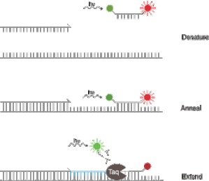
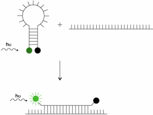
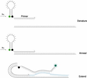
- Introduction define how to distinguish properly a double nearby probe (LC probes), an hydrolysis probe (TaqMan), a molecular beacon and a competitive hybridization probe (Yin-Yang)
- In chapter 3, energy transfer efficiency is detailed. It explains that a nearby quenching system is more efficient than a FRET one. But also that FRET system quenching, having a wider absorption spectrum, quenches more fluorophores than other quenching system.
- At last, many advices are given to do the best choice for the fluorophore/quencher couple considering qPCR device you use (Chapter 4). Here are few of them

   * Considering fluorophore excitation mode (Laser, white light or DEL), excitation efficiency will be different for chose fluorophores. An Argon Laser with blue light will be less efficient for fluorophores having an optimum wavelenght over 540nm vs white light. 
   * If you have single target, you should use fluorophores having a wavelength between 495 and 540nm, that can detected by any qPCR device.
   * If mulitplex qPCR, use fluorophores having the less overlapping spectra, even having a good qPCR device using efficient color compensation.

Another chapter, pretty interesting also, talks about fluorophore quantum yield (efficiency of a fluorophore to convert absorbed light into emitted light). Thus, a high yield gives better fluorescence intensity. But quantum yield can be modified by pH and temperature. Some fluorophore are more sensitive to temperature change. For example, ROX and HEX do not decrease fluorescence up to 85°C whereas FAM decreases steadily but in a moderate way up to 95°C. Cy5 can lose 70% of its quantum yield at 65°C. To offset this effect, probe concentration has to be increased.

Last but not least, it is recalled that a Guanosine base must never be as first probe base and being the closest to grafted fluorophore otherwise the natural quenching effect of G base will affect the fluorophore.
 
This publication has to be read by any qPCR user dealing with qPCR optimization and data analysis. 
Enjoy or in french "Bonne lecture"
 
Reference
Methods Mol Biol. 2006;335:3-16.

Selection of fluorophore and quencher pairs for fluorescent nucleic acid hybridization probes.


With the introduction of simple and relatively inexpensive methods for labeling nucleic acids with nonradioactive labels, doors have been opened that enable nucleic acid hybridization probes to be used for research and development, as well as for clinical diagnostic applications. The use of fluorescent hybridization probes that generate a fluorescence signal only when they bind to their target enables real-time monitoring of nucleic acid amplification assays. The use of hybridization probes that bind to the amplification products in real-time markedly improves the ability to obtain quantitative results. Furthermore, real-time nucleic acid amplification assays can be carried out in sealed tubes, eliminating carryover contamination. Because fluorescent hybridization probes are available in a wide range of colors, multiple hybridization probes, each designed for the detection of a different nucleic acid sequence and each labeled with a differently colored fluorophore, can be added to the same nucleic acid amplification reaction, enabling the development of high-throughput multiplex assays. It is therefore important to carefully select the labels of hybridization probes, based on the type of hybridization probe used in the assay, the number of targets to be detected, and the type of apparatus available to perform the assay. This chapter outlines different aspects of choosing appropriate labels for the different types of fluorescent hybridization probes used with different types of spectrofluorometric thermal cyclers.


mercredi 5 juin 2013

MIQE guidelines : publication or almost a standard?

 I talked about the MIQE guidelines previously, so I am going to give my opinion.
 

MIQE is a publication written by most active scientists about qPCR in the molecular biology planet. Among them, Stephen Bustin, Mike Kubista, Mike Pfaffl, Greg Shipley (great contributors of PCR yahoo forum eg) or Jo Vandesompele.

MIQE is for "Minimum Guidelines for publication of Quantitative real-time PCR Experiments", that is to say a sort of little guide of how is better to publish qPCR results and experiments in order to respect what is a publication or presentation : being able to discuss and reproduce it.

More and more qPCR papers are published and it can be seen from time to time new acronym coming from nowhere or definitions that can be discussed about used techniques being sometimes a bit confusing...

The approach is interesting, but before reading this document, I would like to say that a french standard does exist (followed by other french ones and ISO also) and deal with qPCR : working on Legionella detection but can be read in a second level for all qPCR users because treating results about performances and not only a recipe for Legionella detection only. Named NF T90-471, it has been followed by others like U47-600 working on veterinary samples or ISO 12869, largely inspired from french standard for Legionella.


Back to MIQE.
As said before, the approach has the will to formalize a little bit the exchanges between all qPCR users and also publisher. One of the best reason to me that this text has a real interest was the confusion occuring for a while for the acronym RT-PCR. Indeed, RT would mean "Real-Time"", so quantitative PCR, but also "Reverse Transcription" for PCR using RNA and needing a reverse transcription step to work. This confusion was real for a time among discussion and talks  when people were talking about RT-PCR but thinking about the two different methods. It has lead to real mistake...


This trivial example is not the  main part of MIQE guidelines. This one try to overcome several things like the previous example :

Few things are fixed like :

- "RT-qPCR" for Reverse transcription quantitative PCR and "qPCR" for quantitative PCR.
- "TaqMan probes " are "hydrolysis probe"
- "Housekeeping genes" are"reference genes"
- "FRET probes " are "sondes d'hybridation double"
- "Ct" or "Cp" or "TOP" (not often used) are "Cq" for quantification cycle


Then, a reminder of essential steps to validate a qPCR system is made
:
1 - sensitivity and LOD
2 - specificity
3 - accuracy
4 - repeatibility
5 - reproducibility

PCR is not the only point, another important one is done about getting a good quality sample and standards considering microorganism strain to use, extraction and purification protocol.


Another essential step is the standard quantification. Using spectrophotometry (260/280 ratio) or fluorimetry (with intercalant) is recommended to insure final results and verify what is written on the tube tag which can be wrong or slightly diffierent when we talk about biological smaple.


There are many details in this paper, one is very trivial but I think nowadays not so useless : give all the informations to be able to reproduce experiment in another lab. This can be surprising in a science paper, because it should be the basis of any paper in science. But I agree this has to be reminded because I can quite often read papers where few informations are missing or it is written "following supplier protocol". Except that from time to time, supplier protocol is slightly modified but not written in the paper... and when it is tried to reproduce it, it does not work obviously...as a secret.


In short, this paper is very laudable but is no more than a guide for "how to write a qPCR paper". Is it a sign of any drift?
To conclude, this paper is very useful for anyone wanting to publish or to be able to criticize any results using PCR. But standards are also published and even different, this can lead to dual purpose for several part of the paper between MIQE and qPCR standards. As for me, changing Ct by Cq is not very relevant...


Have a good PCR then and see you next time...

lundi 8 avril 2013

Fluorophores World : Part 2


Now that you have seen a typical qPCR system using a TaqMan probe, let is go to the next step : Duplex.

First of all : why is it better to do a duplex PCR, that is to say having 2 PCR system in the same well with 2 TaqMan probes?

I could answer you briefly without any clear explanation talking about identification, inhibition, control and genotyping. But you will be surely a bit lost and you will not know more about why and how...

One of the main interest for duplex qPCR is : inhibition control

Indeed, for a trivial reason, PCR is an enzymatic reaction prone to inhibition from different compounds. Inhibition has also a major role in the result given. When a positive result is obtained, there is not a lot of question about it, it is taken like that.

On the other hand, when result is negative, does it come from that there is definitely not any target in the sample (true negative) or because the sample is inhibited and PCR does not work well (false negative). How to know?

Using inhibition control is the answer.
This one is composed by a primer and probe system but also by a target DNA. In order to distinguish both in the same well, a difference has to be exploited. The basis fot that is to use a grafted probe with a really different fluorophore from the target one.

Following combination is obtained so :

target DNA
target primers
target probe grafted by FAM for example.

control DNA
control specific primers
control specific probe grafted  with a different probe than the target one


Among available fluorophores, the range is wide and can be seen for example on provider website like   Biosearch or Eurogentec.

The most used ones are FAM (again) and VIC even if it is not the best combination for optimum use, but it is linked to PCR history when these two molecules were the almost only ones on the market, doing good job for duplex. Today, many other exist...

Let is say that we will use FAM and VIC  fluorophores for our duplex example.
It is very important to check fluorophores overlapping for excitation and emission spectra, because it can have a very big impact on detection signal if not well managed. Obviously, FAM and VIC fluorophores have overlapping spectra that has to be compensated then.


These closed spectra induce interferences between the two signals as we would like to discriminate them.
FAM signal (here in green, and one of the strongest emiting fluorophores on the market) overlap the VIC one (in yellow) in a pretty important part. Compared to TAMRA (a lighter overlapping) and Cy5 (no overlap at all), VIC is strongly interfered by FAM. Both signals will be biased and will give bad results for quantification.

To get rid of this problem, a color compensation has to be made. It is a little amplification to do one for all to help the qPCR device to discriminate between both signals. Normally, device supplier can help you if the program is not mentioned in the device notice.
A compensation run comprises a complete mix with two fluorophores and target nucleic acid, but also all the fluorophores in a single detection. When run is done, it will generate a program helping the software device to better discriminate the signals for any detection then.

VIC signal is pretty often the Inhibition Control (IC) signal. And its signal has to be always the same for the Ct. If IC Ct for any sample is the same as the control one, the sample is not considered as inhibited. If the sample Ct IC undergo a Ct shift of at least 3 Ct (1 log), it means that sample is inhibited and that the quantification or detection made is wrong.
If a partial inhibition, IC signal will be shifted and curves flattened, but still positive.
If a complete inhibition, IC signal is not present anymore.

Here a synthesis of results that can be obtained :

Target Control PCR résult Comments
+ + Positive that can be quantified True Positive
+ - Positive with partial inhibition  Partial Inhibition
- + Negative True negative
- - Inhibited Total Inhibition


In the same well, a detection and quantification can be made for a sample but also insuring the results because of inhibition control avoiding true negative.

As told before, many qPCR duplex are made using FAM-VIC, but need color compensation, whereas many other conbination exists working at least the same with less proble to set up (like Cy5-VIC)

In recent years, Quenchers have been improved to replace useful but fluorescent TAMRA one. New quenchers are non-fluorescent and can also help hybridization of target probe like MGB solution from Life Technologies/Applied Biosystem. Multiplex are easier to set up because no backgournd signal coming from emiting quencher like TAMRA.

Most used ones are :

- BHQ (Black Hole Quenchers) I, II et III , depending on the reporter used
- DDQ (Eclipse Dark Quenchers) : similar to BHQ
- MGB (Minor Groove Binder) : molecule improving probe affinity to DNA and comprising also a NFQ (non fluorescent quencher) developed by ABI.

It has to be noticed that using a duplex need quite often working on qPCR parameters to optimize it like IC DNA to use, primers and probe concentrations, hybridization T°, MgCl2 concentration or adjuvant adding.

More details in another post, but also few words on triplex, fluorophores and tips from my PCR experience.

Have good amplifications....

mardi 8 janvier 2013

Fluorophores world : part 1


To understand many things in the qPCR world and to do the right strategic choice, it is better to know few little things about the fantastic world of...fluorophores.

As previously said, PCR systems need primers and if used a probe. I will take a double hybridized probe TaqMan type as an example for this topic. 
These probes are grafted on both extremities by a reporter and a quencher (explained in a previous post)

If using only one probe in the PCR master mix (a simplex), most used fluorophore is then FAM (6-carboyfluorescien or 6-FAM) and the most used quencher is TAMRA (6-carboxy-tetramethyl-rhodamine)

FAM is also one of the most intense molecule considering fluorescence answer to excitation and TAMRA is just the most used for a while but having the drawback to have its own residual fluorescence, able to cause interferences to PCR reaction with small target quantities. TAMRA is replaced more and more by non fluorescent quencher like BHQ (Black Hole Quencher), DDQ (Dark Quencher) or MGB from Applied Biosystem-Life Technologies (comprising a Non fluorescent Quencher).

Hereunder, normalised emission spectra from few fluorophores used in qPCR


It can be observed that FAM and TAMRA spectra are well dissociated and allow a good signal distinction.


In the following table, many more fluorophores are described with their emission and excitation wavelength. Quenchers are also reported on the table side with their use range.


If the table is not clearly visible, here are few links to find any answer about fluorophores association

- IDT report very interesting : http://www.idtdna.com/pages/docs/technical-reports/fluorescence-and-fluorescence-applications.pdf
- Gateway table : http://www.sciencegateway.org/resources/fae1.htm
- Eurogentec guide : http://www.eurogentec.com/uploads/qPCR-guide.pdf

These references are good ones, especially Eurogentec guide. To be honest, I avoid in this blog to repeat one more time things that are described already elsewhere, but sometimes it has to be made. 


We can setup probes having FAM-BHQ1 but not FAM-BHQ3 considering the aborption spectra from the quencher. We can also setup a Cy5-BHQ3 probes but not with BHQ1

Fluorophores choice is essential for a good PCR. It is much more essential when PCR strategy needs a duplex or duplex amplification because chosen ones have to be efficient enough but without having a big wavelength overlapping or too much different fluorescence intensity. 


All of this will be part of part 2.
Have a good PCR.

mardi 16 octobre 2012

Thanks and let's carry on...

Hi all qPCR users (or not),

Just few words to thank all visitors to come and read me because it encourages me to carry on.
To be honest, I do a french version for few years and I decided to translate it for non-french speaking people.
Actually, I have started it because when I wanted to find informations about qPCR, I found few tutorials, forums of course, but almost nothing for troubleshooting, special cases or tips to know about the technique.

And even if you are not thousands of people reading my work, you can be up to hundreds per month to come on my blog.
So I will try to carry on translation and update as fast as possible, but do not hesitate as well to send me questions or comments to make this blog even more interactive.

I write actually about inhibition control and how to handle it with commercial kits but also with personal construction for competitive control to mimic as close as possible target system.

Thanks to everyone and see you soon.

mercredi 3 octobre 2012

Probe strategy : DIY

When the work is done with primers and SybrGreen meaning that PCR answer is good comprising a nice specificity validated with melting curves (single peaks, no shoulders, no close primer-dimer from targeted melting temperatures..), next step is to add a probe to the PCR system and to improve one more time your PCR system.

But talking about probes lead also to talk about different strategies :

First, probes has to be grafted with fluorophores. These molecules has to be wisely chosen depending on your final strategy : simplex (only one target per well) or multiplex (from 2 to 4 different fluorophores in the same well). 4-pex is pretty rare for quantification, we can find some more in genotyping detection.

For example, Eurogentec and Molecular Probes websites provide good table to compare fluorophores spectra you can use for qPCR probes.


In most of the possible strategies about qPCR, there are 2 different molecules called Reporter (fluorescent molecule) and Quencher (molecule that can absorb light energy emitted by Reporter when close enough and  transform it into heat). That is by the way, the FRET principle...
 
When Reporter is excited by a light source, it emits a light but is also immediately quenched and emitted through heat. When the probe is degraded through PCR process, the Reporter is released from the probe, get far from the Quencher and can at least be seen by detection system. 

 
This leads to the following next technology
:

  TaqMan(R) probe
: single oligonucleotide grafted in 5' by the Reporter and in 3' by the Quencher.
This technology relies on Taq polymerase specificity : an exonucleasic activity during complementary strand transcription. Thus, when the probe hybridize on target sequence, there is still no fluorecence detected because Quencher molecule is still close to Reporter. Because reporter is released in the solution due to Taq exonucleasic actitivity and get far from quencher, fluorescence can then be detected.

This method is the most used in qPCR. 


  "FRET"
probes : used mainly by Roche. Actually, this comprises 2 oligonucleotides hybridizing close together. Both are is grafted with one molecule, one is called "donor" and the other one "acceptor". (cf hereunder). Probes place is crucial for a good fluorescence answer. In this method, the two probes are essential to get a signal. When they are well hybridized, they are close enough to operate an energy transfer between them. The donor get the energy and transfer it to acceptor one. Fluorescence will then be detected from this step.


  Molecular Beacon (MB) : 

Another probe type developed by Kramer team in New-York in 1999 
(http://www.molecular-beacons.org/). And because their website is very nice, I will not try to explain MB better than them.

MB design is once again crucial for this specific probe strategie but still using the FRET system for energy transfer. As can be seen on next figure, the probe is grafted with a Reporter (green) and a quencher (black). Its very special hairpin structure needs a very specific design in order to get a specific hybridization without interfering with target hybridization. 
When target sequence hybridizes the probe, this one unfold completely to the sequence. In the meantime, Reporter gets further from the Quencher and fluorescence can occur and be detected. This strategy does not imply hydrolysis of the probe by the Taq and can be used again during nect amplification step. 

This strategy work very well and is used especially for genotyping. Fluorescence has to be detected during hybridization step and not elongation if you use 3-step temperature for PCR reaction. 


Scorpions :

This strategy is more primers modification rather than probe strategy. Somewhere, it is a mix between a MB probe grafted on a primer (see figuer hereunder). Very few references about the use of scorpions primers even if part of many oligo supplier catalog.


Few other strategies exist for qPCR application like Uniprimers, but they are not so used in routine application. 

In a next post, I will talk about fluorophore strategies that can be used on probes. 
Have good amplification and see you next time...